الخميس، 9 أبريل 2020

COVID-19 Attacks the 1-Beta Chain of Hemoglobin

(download pdf file and you will find it also in website) 

The novel coronavirus pneumonia (COVID-19) is an infectious acute respiratory infection caused by the novel coronavirus. 

The virus is a positive-strand RNA virus with high homology to bat coronavirus. 

In this study, conserved domain analysis, homology modeling, and molecular docking were used to compare the biological roles of certain proteins of the novel coronavirus. 

The results showed the ORF8 (open reading frame 8) and surface glycoprotein could bind to the porphyrin, respectively. 

At the same time, orf1ab, ORF10, and ORF3a proteins could coordinate attack the heme on the 1-beta chain of hemoglobin to dissociate the iron to form the porphyrin. 

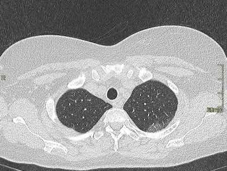
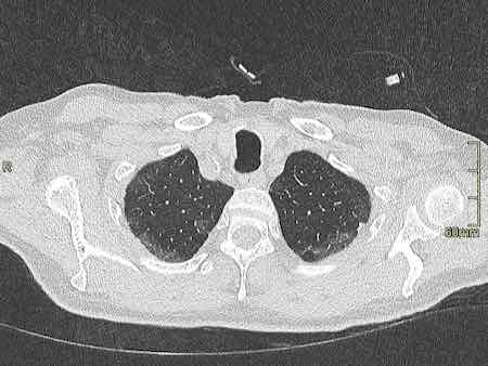
The attack will cause less and less hemoglobin that can carry oxygen and carbon dioxide. The lung cells have extremely intense poisoning and inflammatory due to the inability to exchange carbon dioxide and oxygen frequently, which eventually results in ground-glass-like lung images. 

The mechanism also interfered with the normal heme anabolic pathway of the human body, is expected to result in human disease. According to the validation analysis of these finds, chloroquine could prevent orf1ab, ORF3a, and ORF10 to attack the heme to form the porphyrin, and inhibit the binding of ORF8 and surface glycoproteins to porphyrins to a certain extent, effectively relieve the symptoms of respiratory distress. 

Favipiravir could inhibit the envelope protein and ORF7a protein bind to porphyrin, prevent the virus from entering host cells, and catching free porphyrins. 

Because the novel coronavirus is dependent on porphyrins, it may originate from an ancient virus. Therefore, this research is of high value to contemporary biological experiments, disease prevention, and clinical treatment.

Covid-19 no pnemumonia or ARSD

Covid-19 had us all fooled, but now we might have finally found its secret.

In the last 3–5 days, a mountain of anecdotal evidence has come out of NYC, Italy, Spain, etc. about COVID-19 and characteristics of patients who get seriously ill. It’s not only piling up but now leading to a general field-level consensus backed up by a few previously little-known studies that we’ve had it all wrong the whole time. Well, a few had some things eerily correct (cough Trump cough), especially with Hydroxychloroquine with Azithromicin, but we’ll get to that in a minute.
There is no ‘pneumonia’ nor ARDS. At least not the ARDS with established treatment protocols and procedures we’re familiar with. Ventilators are not only the wrong solution, but high pressure intubation can actually wind up causing more damage than without, not to mention complications from tracheal scarring and ulcers given the duration of intubation often required… They may still have a use in the immediate future for patients too far to bring back with this newfound knowledge, but moving forward a new treatment protocol needs to be established so we stop treating patients for the wrong disease.
The past 48 hours or so have seen a huge revelation: COVID-19 causes prolonged and progressive hypoxia (starving your body of oxygen) by binding to the heme groups in hemoglobin in your red blood cells. People are simply desaturating (losing o2 in their blood), and that’s what eventually leads to organ failures that kill them, not any form of ARDS or pneumonia. All the damage to the lungs you see in CT scans are from the release of oxidative iron from the hemes, this overwhelms the natural defenses against pulmonary oxidative stress and causes that nice, always-bilateral ground glass opacity in the lungs. Patients returning for re-hospitalization days or weeks after recovery suffering from apparent delayed post-hypoxic leukoencephalopathy strengthen the notion COVID-19 patients are suffering from hypoxia despite no signs of respiratory ‘tire out’ or fatigue.
Here’s the breakdown of the whole process, including some ELI5-level cliff notes. Much has been simplified just to keep it digestible and layman-friendly.
Your red blood cells carry oxygen from your lungs to all your organs and the rest of your body. Red blood cells can do this thanks to hemoglobin, which is a protein consisting of four “hemes”. Hemes have a special kind of iron ion, which is normally quite toxic in its free form, locked away in its center with a porphyrin acting as it’s ‘container’. In this way, the iron ion can be ‘caged’ and carried around safely by the hemoglobin, but used to bind to oxygen when it gets to your lungs.
When the red blood cell gets to the alveoli, or the little sacs in your lungs where all the gas exchange happens, that special little iron ion can flip between FE2+ and FE3+ states with electron exchange and bond to some oxygen, then it goes off on its little merry way to deliver o2 elsewhere.
Here’s where COVID-19 comes in. Its glycoproteins bond to the heme, and in doing so that special and toxic oxidative iron ion is “disassociated” (released). It’s basically let out of the cage and now freely roaming around on its own. This is bad for two reasons:
1) Without the iron ion, hemoglobin can no longer bind to oxygen. Once all the hemoglobin is impaired, the red blood cell is essentially turned into a Freightliner truck cab with no trailer and no ability to store its cargo.. it is useless and just running around with COVID-19 virus attached to its porphyrin. All these useless trucks running around not delivering oxygen is what starts to lead to desaturation, or watching the patient’s spo2 levels drop. It is INCORRECT to assume traditional ARDS and in doing so, you’re treating the WRONG DISEASE. Think of it a lot like carbon monoxide poisoning, in which CO is bound to the hemoglobin, making it unable to carry oxygen. In those cases, ventilators aren’t treating the root cause; the patient’s lungs aren’t ‘tiring out’, they’re pumping just fine. The red blood cells just can’t carry o2, end of story. Only in this case, unlike CO poisoning in which eventually the CO can break off, the affected hemoglobin is permanently stripped of its ability to carry o2 because it has lost its iron ion. The body compensates for this lack of o2 carrying capacity and deliveries by having your kidneys release hormones like erythropoietin, which tell your bone marrow factories to ramp up production on new red blood cells with freshly made and fully functioning hemoglobin. This is the reason you find elevated hemoglobin and decreased blood oxygen saturation as one of the 3 primary indicators of whether the shit is about to hit the fan for a particular patient or not.
2) That little iron ion, along with millions of its friends released from other hemes, are now floating through your blood freely. As I mentioned before, this type of iron ion is highly reactive and causes oxidative damage. It turns out that this happens to a limited extent naturally in our bodies and we have cleanup & defense mechanisms to keep the balance. The lungs, in particular, have 3 primary defenses to maintain “iron homeostasis”, 2 of which are in the alveoli, those little sacs in your lungs we talked about earlier. The first of the two are little macrophages that roam around and scavenge up any free radicals like this oxidative iron. The second is a lining on the walls (called the epithelial surface) which has a thin layer of fluid packed with high levels of antioxidant molecules.. things like abscorbic acid (AKA Vitamin C) among others. Well, this is usually good enough for naturally occurring rogue iron ions but with COVID-19 running rampant your body is now basically like a progressive state letting out all the prisoners out of the prisons… it’s just too much iron and it begins to overwhelm your lungs’ countermeasures, and thus begins the process of pulmonary oxidative stress. This leads to damage and inflammation, which leads to all that nasty stuff and damage you see in CT scans of COVID-19 patient lungs. Ever noticed how it’s always bilateral? (both lungs at the same time) Pneumonia rarely ever does that, but COVID-19 does… EVERY. SINGLE. TIME.
— — — — — — — — — — — — -
Once your body is now running out of control, with all your oxygen trucks running around without any freight, and tons of this toxic form of iron floating around in your bloodstream, other defenses kick in. While your lungs are busy with all this oxidative stress they can’t handle, and your organs are being starved of o2 without their constant stream of deliveries from red blood cell’s hemoglobin, and your liver is attempting to do its best to remove the iron and store it in its ‘iron vault’. Only its getting overwhelmed too. It’s starved for oxygen and fighting a losing battle from all your hemoglobin letting its iron free, and starts crying out “help, I’m taking damage!” by releasing an enzyme called alanine aminotransferase (ALT). BOOM, there is your second of 3 primary indicators of whether the shit is about to hit the fan for a particular patient or not.
Eventually, if the patient’s immune system doesn’t fight off the virus in time before their blood oxygen saturation drops too low, ventilator or no ventilator, organs start shutting down. No fuel, no work. The only way to even try to keep them going is max oxygen, even a hyperbaric chamber if one is available on 100% oxygen at multiple atmospheres of pressure, just to give what’s left of their functioning hemoglobin a chance to carry enough o2 to the organs and keep them alive. Yeah we don’t have nearly enough of those chambers, so some fresh red blood cells with normal hemoglobin in the form of a transfusion will have to do.
The core point being, treating patients with the iron ions stripped from their hemoglobin (rendering it abnormally nonfunctional) with ventilator intubation is futile, unless you’re just hoping the patient’s immune system will work its magic in time. The root of the illness needs to be addressed.
Best case scenario? Treatment regimen early, before symptoms progress too far. Hydroxychloroquine (more on that in a minute, I promise) with Azithromicin has shown fantastic, albeit critics keep mentioning ‘anecdotal’ to describe the mountain, promise and I’ll explain why it does so well next. But forget straight-up plasma with antibodies, that might work early but if the patient is too far gone they’ll need more. They’ll need all the blood: antibodies and red blood cells. No help in sending over a detachment of ammunition to a soldier already unconscious and bleeding out on the battlefield, you need to send that ammo along with some hemoglobin-stimulant-magic so that he can wake up and fire those shots at the enemy.

The story with Hydroxychloroquine

All that hilariously misguided and counterproductive criticism the media piled on chloroquine (purely for political reasons) as a viable treatment will now go down as the biggest Fake News blunder to rule them all. The media actively engaged their activism to fight ‘bad orange man’ at the cost of thousands of lives. Shame on them.
How does chloroquine work? Same way as it does for malaria. You see, malaria is this little parasite that enters the red blood cells and starts eating hemoglobin as its food source. The reason chloroquine works for malaria is the same reason it works for COVID-19 — while not fully understood, it is suspected to bind to DNA and interfere with the ability to work magic on hemoglobin. The same mechanism that stops malaria from getting its hands on hemoglobin and gobbling it up seems to do the same to COVID-19 (essentially little snippets of DNA in an envelope) from binding to it. On top of that, Hydroxychloroquine (an advanced descendant of regular old chloroquine) lowers the pH which can interfere with the replication of the virus. Again, while the full details are not known, the entire premise of this potentially ‘game changing’ treatment is to prevent hemoglobin from being interfered with, whether due to malaria or COVID-19.
No longer can the media and armchair pseudo-physicians sit in their little ivory towers, proclaiming “DUR so stoopid, malaria is bacteria, COVID-19 is virus, anti-bacteria drug no work on virus!”. They never got the memo that a drug doesn’t need to directly act on the pathogen to be effective. Sometimes it’s enough just to stop it from doing what it does to hemoglobin, regardless of the means it uses to do so.
Anyway, enough of the rant. What’s the end result here? First, the ventilator emergency needs to be re-examined. If you’re putting a patient on a ventilator because they’re going into a coma and need mechanical breathing to stay alive, okay we get it. Give ’em time for their immune systems to pull through. But if they’re conscious, alert, compliant — keep them on O2. Max it if you have to. If you HAVE to inevitably ventilate, do it at low pressure but max O2. Don’t tear up their lungs with max PEEP, you’re doing more harm to the patient because you’re treating the wrong disease.
Ideally, some form of treatment needs to happen to:
  1. Inhibit viral growth and replication. Here plays CHQ+ZPAK+ZINC or other retroviral therapies being studies. Less virus, less hemoglobin losing its iron, less severity and damage.
  2. Therapies used for anyone with abnormal hemoglobin or malfunctioning red blood cells. Blood transfusions. Whatever, I don’t know the full breadth and scope because I’m not a physician. But think along those lines, and treat the real disease. If you’re thinking about giving them plasma with antibodies, maybe if they’re already in bad shape think again and give them BLOOD with antibodies, or at least blood followed by plasma with antibodies.
  3. Now that we know more about how this virus works and affects our bodies, a whole range of options should open up.
  4. Don’t trust China. China is ASSHOE. (disclaimer: not talking about the people, just talking about the regime). They covered this up and have caused all kinds of death and carnage, both literal and economic. The ripples of this pandemic will be felt for decades.
السبت، 4 أبريل 2020

COVID-19 Imaging findings (from radiologyassistant)


Chest CT


Ground glass

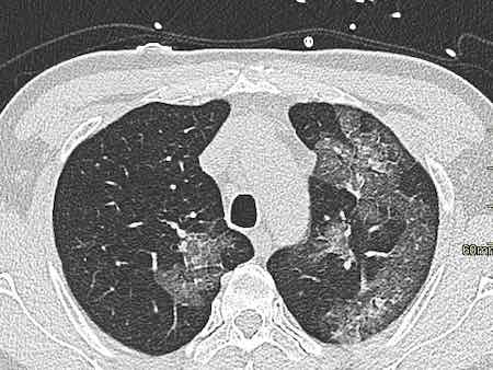
Ground glass (GGO) pattern is the most common finding in COVID-19 infections.
They are usually multifocal, bilateral and peripheral, but in the early phase of the disease the GGO may present as a unifocal lesion, most commonly located in the inferior lobe of the right lung (6).
CT-images of a young male, who had fever for ten days with progressive coughing and shortness of breath.
Saturation at admission was 66%.
The PCR test was positive for COVID-19.
There are widespread bilateral ground-glass opacities with a posterior predominance.

Crazy paving

Sometimes there are thickened interlobular and intralobular lines in combination with a ground glass pattern.
This is called crazy paving.
It is believed that this pattern is seen in a somewhat later stage.

Traction Bronchiectasis

Another common finding in the areas of ground glass is traction bronchiectasis (arrows).

Subpleural bands and Architectural distortion

In some case there is architectural distortion with the formation of subpleural bands.

COVID-19 infection. Predominantly bilateral subpleural GGO with some areas of crazy paving. In the lower lobes some areas of consolidation. Percentage of lung involvement is approximately 25% by visual assessment.

CT involvement score

The severity of the lung involvement on the CT correlates with the severity of the disease.
Visual assessment
The severity on CT can be estimated by visual assessment.
This is the easiest way to score the severity.
The CT images show a 25% involvement by visual assessment.
Severity score
Another method is by scoring the percentages of each of the five lobes that is involved:
  1. < 5% involvement
  2. 5%-25% involvement
  3. 26%-49% involvement
  4. 50%-75% involvement 
  5. > 75% involvement. 
The total CT score is the sum of the individual lobar scores and can range from 0 (no involvement) to 25 (maximum involvement), when all the five lobes show more than 75% involvement.
Some say that the percentage of lung involvement can be calculated by multiplying the total score times 4.
This however is not true. Suppose that all lobes have a 10% involvement, then this would lead to an overall score of 10, which could lead to the impression that 40% of the lungs are involved.
Common Patterns and Distribution on Initial CT Images of 919 patients COVID-19 (4).

Initial CT-findings

Initial CT-findings in COVID-19 cases include bilateral, multilobar groud glass opacification (GGO) with a peripheral or posterior distribution, mainly in the lower lobes nd less frequently in the middle lobe (4).
Consolidation superimposed on GGO as the initial imaging presentation is found in a smaller number of cases, mainly in the elderly population.
Septal thickening, bronchiectasis, pleural thickening, and subpleural involvement are some of the less common findings, mainly in the later stages of the dis- ease.
Pleural effusion, pericardial effusion, lymphadenopathy, cavitation, CT halo sign, and pneumothorax are some of the uncommon but possible findings seen with disease progression.
There is much overlap of the CT-pattern of COVID-19 with other viral pneumonias.

CT-images of a 78 year old male with coughing for 2 weeks and progressive shorteness of breath, who tested positive for COVID-19.
There are bilateral ground-glass opacities with a posterior predominance.
In the right lower lobe there is a consolidation and there is bronchiectasis especially in the left lower lobe.
About 75% of the lungs are involved.
Click to view larger image.
Images of a 59 year old male who had fever for one week with non-productive cough.
The PCR-test was negative.
Because of clinical suspicion a CT was performed which showed some areas of GGO and massive consolidation in the posterior parts of the lower lobes (arrow on sagittal reconstruction).
Two days later a sputum test was positive for COVID-19.

Changes over time

Advanced-phase disease is associated with a significantly increased frequency of:
  • GGO plus a reticular pattern (crazy pavin)
  • Vacuolar sign
  • Fibrotic streaks
  • Air bronchogram
  • Bronchus distortion
  • Subpleural line or a subpleural transparent line 
  • Pleural effusion
Early phase COVID-19
This 59 year old female had a history of ten days of fever and five days of coughing.
The O2 saturation was 89 and her respiratory rate was 30/min (normal: 12-18).
There are widespread GGO's without consolidation. No architectural distortion.
This was reported as early phase COVID-19.
These images are of a 49 year old male with fever, cough and a low saturation.
The images show:
  • Bilateral areas of GGO.
    The ground glass density is more pronounced
  • Fibrotic bands (arrows).
  • Dilated vessels in affected area (circle).
Based on the CT-findings COVID-19 infection was assumed to be highly likely - late phase.
COVID-19 infection - late phase
This patient had fever for one week with some abdominal pain and diarrhoea.
On the day of admission she had a dry cough and complained of dizziness.
The O2-saturation was low.
The PCR-test was not known and a CT was performed for triage.
The images show:
  • Bilateral subpleural GGO's 
  • Consolidation in right lower lobe with traction bronchiectasis (green arrow).
  • Fibrous bands (yellow arrow).
Based on the CT-findings COVID-19 infection was assumed to be highly likely - late phase.

CT Report

In the tabel a checklist of CT findings to mention in the report.
In the first four days after the presentation of the complaints, the CT is not sensitive as initial test as 50% of patients may have a normal CT.
After these first four days, the CT has a very high sensitivity.

Chest radiograph

Courtesy Dr. Michael David Kuo (1).
The chest film is insensitive early in the disease.
Here a comparison of a chest radiograph and CT image.
The ground glass opacities in the right lower lobe on the CT (red arrows) are not visible on the chest radiograph, which was taken 1 hour prior to the CT-study (1). 
Chest-films can be useful in the follow-up of the disease.
These x-rays are of a patient with COVID-19.
On admission to the hospital the chest film was normal.
Four days later the patient is on mechanical ventilation and there are bilateral consolidations on the chest film.
Chest film of a 83 year old male with mitral insufficiency, pulmonary hypertension and atrial fibrillation with COVID-19 infection.
Ground-glass opacification and consolidation in right upper lobe and left lower lobe (arrows).
Courtesy of Dr. Edgar Lorente (5)
A series of chest films of a 72-year-old woman admitted with acute respiratory failure, fever (38ºC) and dyspnoea.
She was tachypneic (30bpm), with lymphopenia and low oxygen saturation (SpO2 85%).
Patient presented to the emergency department two days earlier with fever (up to 38.6ºC), dry cough, odynophagia and general malaise.
She was discharged from hospital because she did not present alarm criteria at that time.
The patient required mechanical ventilation and was admitted to intensive care.
During her stay in ICU, poor evolution to respiratory distress syndrome and to multi-organic failure.
The patient died 24 hours later.
Imaging findings:
  • At admission: Ill-defined bilateral alveolar consolidation with peripheral distribution.
  • 4 hours later: Radiological worsening, with affectation of lower lobes. Endotracheal tube and central venous line were required.
  • 24 hours: Bilateral alveolar consolidation.
  • 48 hours: Radiological worsening. Bilateral alveolar consolidation with panlobar affectation.
  • 72 hours: Bilateral alveolar consolidation with panlobar affectation, with typical radiological findings of ARDS. 24 hours later the patient passed away.

Possible role of CT

CT can play a role in:
  • Triage of patients:
    - no COVID-19
    - possible or most likely COVID-19
    - severity of the disease
  • Prediction of worsening
  • Prediction of improvement
  • Problem solver
Triage
Some published clinical guidelines recommend chest CT for patients with suspected COVID-19.
The decision to use of CT for triage depends on many considerations:
  • a priori chance of COVID-19 infection.
  • CT availability, for instance can one CT be used as Corona-CT or is there a CT near the emergency room.
  • clinical suspicion in patients with negative PCR.

Video cases of CT Chest

...
This is a 67 year old woman who was coughing for one week and now presented with shortness of breath.
This is a CT-scan of a 53 year old woman at admission.
There are 2 areas with a halo sign, some areas of ground-glass and consolidations in the lower lobes.
Based on the CT-findings she was suspected of having COVID-19.
The PCR-test the next day was positive.
60 year old male with complaints of fatigue and coughing for one week.

Typical and Atypical symptoms of COVID-19

Typical and Atypical symptoms of COVID-19


***
Corona family: (To remember)
Wuhan Novel Coronavirus (2019-nCoV)
SARS
MERS
Swine Flu
***
1- many people have no symptoms at all.
2- most common distinctive symptoms of COVID-19 are: 
           a dry cough, 
           fever, and 
           difficulty breathing.

3- nausea and diarrhea, abdominal pain  (see this website)
(up to half of patients may have digestive issues along with respiratory symptoms)

4- loss of smell and taste

5- malaise, a general sense of discomfort, illness, or uneasiness

6- disorientation, or exhaustion

7- Chills or muscle aches

8- Headaches and dizziness

*******
Clinical manifestations (uptodate)
Initial presentation — Pneumonia appears to be the most frequent serious manifestation of infection, characterized primarily by fever, cough, dyspnea, and bilateral infiltrates on chest imaging. There are no specific clinical features that can yet reliably distinguish COVID-19 from other viral respiratory infections.
In a study describing 138 patients with COVID-19 pneumonia in Wuhan, the most common clinical features at the onset of illness were:
       Fever in 99 percent
Fatigue in 70 percent

Dry cough in 59 percent

Anorexia in 40 percent

Myalgias in 35 percent

Dyspnea in 31 percent

Sputum production in 27 percent
Other cohort studies of patients from Wuhan with confirmed COVID-19 have reported a similar range of clinical findings. However, fever might not be a universal finding. 
In one study, fever was reported in almost all patients, but approximately 20 percent had a very low grade fever <100.4°F/38°C. 
In another study of 1099 patients from Wuhan and other areas in China, fever (defined as an axillary temperature over 99.5°F/37.5°C) was present in only 44 percent on admission but was ultimately noted in 89 percent during the hospitalization .
Although not highlighted in the initial cohort studies from China, smell and taste disorders (eg, anosmia and dysgeusia = is a distortion of the sense of taste) have also been reported as common symptoms in patients with COVID-19
In a survey of 59 patients with COVID-19 in Italy, 34 percent self-reported either a smell or taste aberration and 19 percent reported both . Whether this is a distinguishing feature of COVID-19 is uncertain.
Other, less common symptoms have included headache, sore throat, and rhinorrhea. In addition to respiratory symptoms, gastrointestinal symptoms (eg, nausea and diarrhea) have also been reported; and in some patients, they may be the presenting complaint.
.